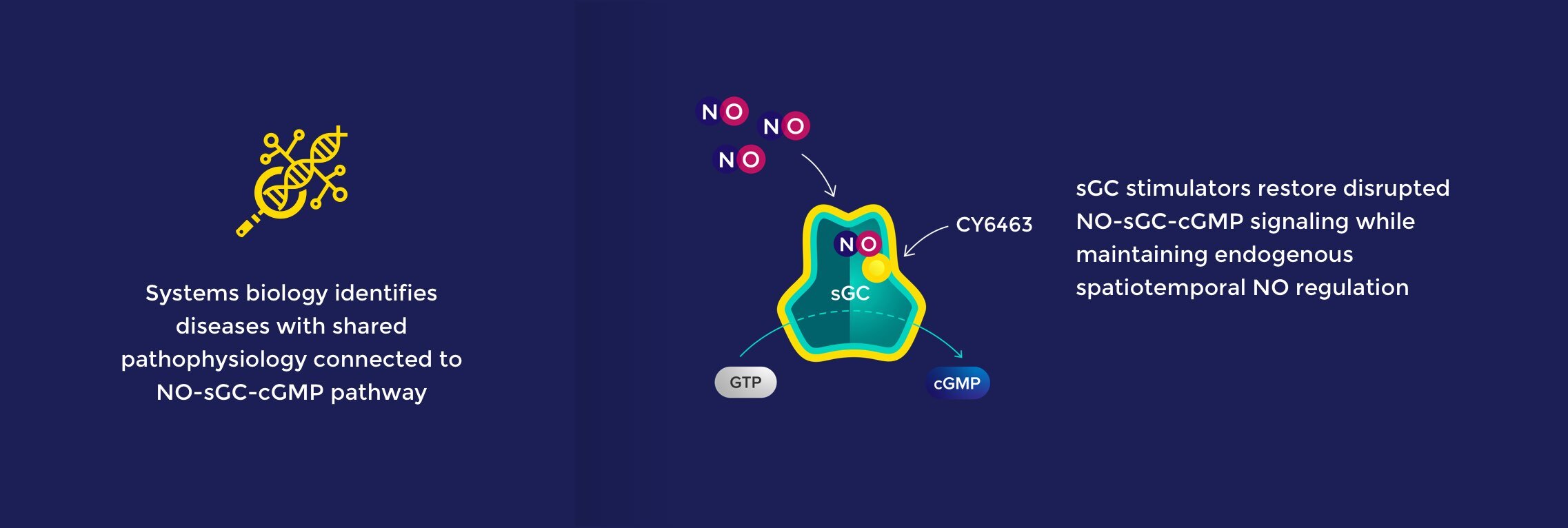
CNS

CNS is a high reward space where the large unmet needs are growing, given aging patient populations and the dearth of approved and effective treatments.

NO-sGC-cGMP and Brain Health

In the CNS, NO-sGC-cGMP signaling underlies multiple physiological processes that contribute to overall brain health, including neurotransmission, neurovascular function, cellular bioenergetics, and inflammation. Nitric oxide is a gas with a short half-life (0.5-5s) that acts locally (100-200μm range). These properties enable the precise and dynamic spatial and temporal regulation seen with NO and other neurotransmitters.

NO-sGC-cGMP signaling underlies multiple physiological
processes that contribute to overall brain health

Our systems biology approach combines genetic and proteomic data to identify CNS diseases in which the NO-sGC-cGMP pathway plays a central role. This approach identified a broad range of both neurodegenerative diseases (e.g., Alzheimer’s Disease) and neuropsychiatric diseases (e.g., schizophrenia) – with dysfunctional NO-sGC-cGMP signaling as the common pathophysiologic thread.

sGC stimulators, like CY6463, are allosteric modulators of sGC and amplify endogenous NO signaling. In diseases associated with deficits in the NO-sGC-cGMP pathway, CY6463 has the potential to address the underlying pathophysiology by restoring appropriate endogenous signaling and maintaining the precise spatial and temporal control that is the hallmark of this signaling pathway.

CY6463 is the first CNS-penetrant sGC stimulator designed and in development for neurodegenerative diseases. In preclinical studies, CY6463 showed benefits in multiple animal models and across four domains of human neurodegenerative diseases.

Preclinical data demonstrate sGC stimulator effects across multiple CNS domains

Improves

Neuronal
Function

Enhanced memory performance & spine density in aged animals; increased LTP in neurodegenerative disease models

Reduces

Neuro-
inflammation

Decreased markers of LPS-induced neuroinflammation (ICAM1, VCAM1, IL6) in vitro

Enhances

Cellular
Bioenergetics

Increased ATP and restored gene expression in cells from patients with mitochondrial diseases

Improves

Cerebral
Blood Flow

Increased blood flow in areas associated with memory and arousal by fMRI BOLD imaging

CNS Opportunities

CNS is a high-reward space with large and growing unmet need due to an aging population and a dearth of approved and effective treatments. Rapidly evolving science, including both genetic insights and technologies, is accelerating the discovery and development of new CNS drugs.

The sGC mechanism has the potential to create a meaningful medical breakthrough by stimulating a previously undrugged neurotransmitter system in the CNS. Other successfully drugged neurotransmitter systems opened entirely new therapeutic categories and created significant market opportunities. This type of therapeutic potential creates broad opportunity to help a wide range of patients suffering from CNS diseases, and thus makes our CNS program an exciting strategic focus today and a core part of Cyclerion going forward.

CNS Reference Publications

NO-sGC-cGMP signaling in the CNS 

  • Garthwaite, John. “Nitric oxide as a multimodal brain transmitter.” Brain and neuroscience advances vol. 2 2398212818810683. 4 Dec. 2018
    This review outlines the discovery and growing understanding of nitric oxide (NO) as a brain transmitter and soluble guanylate cyclase (sGC) as its primary receptor, including a description of the synaptic location of sGC and the NO-sGC binding and conformational changes that lead to cGMP production. The author notes that NO operates across brain regions and is involved in learning and memory formation, moving, feeding, sleeping, reproduction, pain and anxiety, brain development and synaptogenesis. This broad role reflects the nature of NO, which is a versatile transmitter. NO can act in different ways depending on its source (NOS) location, which is largely but not exclusively in neurons and can be pre- or postsynaptic, and its receptor location, which can be postsynaptic or presynaptic in neurons, in astrocytes, oligodendrocytes, and vascular cells. Three modes of NO signaling are described that differ in the reach of their communication (single synapse, synaptic spillover, and volume transmission) and are determined by differences in the location(s) of the NO source(s) and the rate/volume of NO production across single to multiple sources.
  • Kleppisch T, Feil R. cGMP signalling in the mammalian brain: role in synaptic plasticity and behaviour. Handb Exp Pharmacol. 2009;(191):549-79.
    This review (co-authored by our collaborator Dr. Feil) summarizes biochemical and functional data on the role of the cGMP signaling pathway in the mammalian brain, with a focus on the regulation of synaptic plasticity, learning, and other complex behaviors. The expression and role of central players in the pathway (including NOS-NosGC-cGMP-PKG-PDE)  in the specific areas of the CNS, as well as downstream targets relevant for pre- and post-synaptic CNS function (including CNG, HCN, AMPA, NMDAR, CREB) are discussed.
  • Ben Aissa M, Lee SH, Bennett BM, Thatcher GR. Targeting NO/cGMP Signaling in the CNS for Neurodegeneration and Alzheimer’s Disease. Curr Med Chem. 2016;23(24):2770-2788.
    This review focuses on CREB as a relevant target of NO-sGC-cGMP signaling in the CNS. The central role of CREB in synaptic plasticity is described, as is the role of NO-cGMP in neurodegeneration. Disease-modifying pharmacological approaches to treat cognition and memory dysfunction in neurodegenerative diseases (with a focus on AD) by targeting the NO-sGC pathway (with NO donors, sGC stimulators, PDE inhibitors) are discussed.
  • Hollas MA, Ben Aissa M, Lee SH, Gordon-Blake JM, Thatcher GRJ. Pharmacological manipulation of cGMP and NO/cGMP in CNS drug discovery. Nitric Oxide. 2019 Jan 1;82:59-74.
    This review highlights the central role played by NO-cGMP in learning and memory, and the substantial evidence that the NO-cGMP signaling pathway is perturbed in neurodegenerative disorders (including AD). The disease-modifying effects of NO-chimeras/nitrates and PDE inhibitors in preclinical animal models are discussed, as are the downstream mechanisms (eg, via GABAA and NMDA receptors) mediating these effects on synaptic and neuronal dysfunction. Finally, the more recently recognized potential for allosteric modulation of sGC in the brain is described, especially in light of the lack of success of PDE inhibitors in clinical trials of brain disorders.

qEEG spectral frequency analysis

  • Ishii et al. Healthy and Pathological Brain Aging: From the Perspective of Oscillations, Functional Connectivity, and Signal Complexity. Neuropsychobiology, 2018
    This comprehensive review of neurocognitive aging from a neurophysiological perspective highlights the value of EEG in understanding physiological mechanisms associated with brain aging. For example, EEG is noninvasive and measures brain electromagnetic activity with high temporal resolution (ms). Physiological and pathological aging affects EEG alpha rhythm (refer to Table 1 in paper) including reductions in amplitude of resting alpha, decrease in posterior cortical alpha magnitude, decrease in occipital lobe alpha power, and decrease in alpha rhythm reactivity. These age-related findings in alpha, as well as other EEG changes, have been variously linked to poor cognitive performance (including memory performance), genetic factors (ie, CLU and APOE4), hippocampal and cortical atrophy, AD and AD biomarkers (Aβ42 and tau), and brain network organization. Rich reference list.
  • Babiloni, et al. Resting-state posterior alpha rhythms are abnormal in subjective memory complaint seniors with preclinical Alzheimer’s neuropathology and high education level: the INSIGHT-preAD study. Neurobiol Aging. 2020;90:43-59.
    Cognitive reserve represents an individual’s resilience in maintaining cognitive function despite age- or disease-related neuropathic and neurodegenerative changes as measured by markers in CSF, PET tracers, MRI, etc. Older adults with high educational attainment, lifespan intellectual activity, etc., demonstrate cognitive reserve, which may confer neuroprotective and compensatory mechanisms. qEEG spectral analysis may offer a noninvasive, low-cost alternative to expensive and invasive neuroimaging for monitoring neuropathy and neurodegeneration; abnormalities in posterior alpha rhythms with ADMCI, AD, and MCI due to Parkinson’s and Lewy Body disease have been demonstrated. In this retrospective study, the authors tested the hypothesis that subjects with subjective memory complaint with preclinical AD pathology (SMCpos) (as demonstrated by PET) and high education level may show abnormalities in resting state posterior alpha rhythms (quiet wakefulness) consistent with the idea of cognitive reserve. The study demonstrated that SMCneg seniors with high (relative to low) education level had higher power density (amplitude) posterior alpha rhythms (possibly neuroprotective). SMCpos with high education showed higher power temporal alpha (possibly neuroprotective) and lower power posterior alpha rhythms (possibly compensatory). These differences may represent cognitive reserve as subjects had no differences in cognitive function or brain gray-white matter.
  • Marin et al. Peak alpha frequency and N200 latency as predictors of neuropsychological performance in a memory disorders clinic. Alzheimer’s Association International Conference 2021.
    This poster from the laboratory of Dr. Andrew Budson and Dr. Katherine Turk in the Department of Neurology, Boston University School of Medicine and Center for Translational and Cognitive Neuroscience, VA Boston Healthcare System is a Cyclerion sponsored study on peak alpha frequency and N200 latency as predictors of neuropsychological performance in a memory disorders clinic. This pioneering work provides insights into the relationships between electrophysiological measures and cognitive performance in patients with Alzheimer’s disease and other dementias.

Event-related potential (ERP): N200 and P300

  • Bennys K, Portet F, Touchon J. Diagnostic value of event-related evoked potentials N200 and P300 subcomponents in early diagnosis of Alzheimer’s disease and mild cognitive impairment. J Clin Neurophysiol 2007;24:405–12.
    The introduction concisely describes how ERP study conditions relate to cognitive phenomena and how the N200 and P300 components are thought to relate to specific cognitive processes. For example, N200 may be linked to the detection of the target stimulus and be the reflection of the selective attention processes coming into action. This study assessed N200 and P300 (ERP oddball paradigm) in 3 groups: 30 mild to moderate potential AD, 20 MCI, and 10 healthy subjects. N200 and P300 latencies differentiated AD from MCI, AD from healthy, and MCI from healthy; P300 latency was 100% specific to AD vs MCI. Figure 2 shows latencies in the 3 groups at different regions. N200 and P300 latencies in frontal and parietal regions negatively correlated with MMSE and positively correlated with executive and attention functions (highest latency, lowest performance). The authors conclude: “These noninvasive examinations are able to point at an early stage an alteration of the cognitive functions and can contribute to the diagnosis for Alzheimer’s disease with both good sensitivity and specificity.”
  • Fruehwirt et al. Associations of event-related brain potentials and Alzheimer’s disease severity: A longitudinal study. Progress in Neuropsychopharmacology and Biological Psychiatry 92 (2019) 31-38.
    There are no cost-efficient biomarkers for the diagnosis of early AD in clinical practice. This study investigated the correlation of ERP P300, N200, and P50 (oddball paradigm) with AD severity by MMSE at baseline (BL) and then followed the ERP components for 18m in 63 subjects [31 possible AD, 32 probable AD; 39 ApoE ɛ4 allele carriers; 39 on anti-dementia treatment (acetylcholinesterase inhibitors, NMDA receptor antagonists); 38 females; mean age 75.92y]. P300 and N200 latency correlated with disease severity in the group of all AD patients, as well as the subgroup of probable AD patients at BL, whereas P50 amplitude did not show significant correlations in any group. P300 latency, which showed the strongest association with MMSE at BL, significantly increased over the 18m but the change did not correlate with MMSE score change.

Saccadic eye movement (SEM)

  • Wilcockson et al. Abnormalities of saccadic eye movements in dementia due to Alzheimer’s disease and mild cognitive impairment. Aging 2019, Vol.11, No.15.
    68 AD subjects, 42 with amnesic MCI (aMCI), 47 with non-amnesic MCI (naMCI), and 92 healthy elderly subjects completed antisaccade (AS) and cognitive testing.  The objective of the study was to determine whether SEM could distinguish between aMCI and naMCI subjects, since aMCI subjects are at greater risk of conversion to AD.  AS latencies and AS errors for AD and aMCI were greater than naMCI and controls, but AD and aMCI were not different and naMCI and controls were not different.  The AS error rate was found to correlate with the FCSRT free recall score, where increasing errors were associated with poorer cognitive performance.  Therefore, the authors conclude that AS measures can distinguish aMCI from naMCI and have potential as a biomarker for dementia.
  • James A. Sharpe & David H. Zackon (1987) Senescent Saccades: Effects of Aging on Their Accuracy, Latency and Velocity, Acta Oto-Laryngologica, 104:5-6, 422-428
    12 young, 11 middle-aged, and 11 elderly normal subjects completed SEM tasks involving stimuli with 1. Predictable amplitude, directions, and timing, 2. Unpredictable amplitudes and directions at regular intervals, or 3. Unpredictable timing of predictable amplitude and direction.  Latencies were greater in elderly subjects for all testing types.  Peak velocities were reduced in elderly when target amplitude and direction were predictable (conditions 1 and 3).  Elderly subjects also had increased errors (conditions 1 and 2).  In the discussion, the authors speculate about cell types, brain regions, and aspects of cognition that may be involved in explaining their findings.  In conclusion, aging does impact SEM measures and should be considered as a variable.
Top